Mechanobiology of Palatal Shelf Elevation
Student: Pending
Investigators: Ken Fischer, PhD; Irfan Saadi, PhD
Clefts of palate are the most common craniofacial anomalies among birth defects. Orofacial clefts occur as part of >400 syndromes or as isolated cases. Isolated or non-syndromic cleft palate (CP) with or without cleft lip alone occur in approximately 1/1200 births worldwide and have a complex etiology, including both genetic and environmental (biochemical and biomechanical) factors, that is poorly understood. Although the anatomy and biology of palate development has been extensively studied, the mechanisms underlying vertical to horizontal palate elevation remain enigmatic. This research is developing pioneering FEA models of embryonic palate tissues and determine appropriate material models and material properties. We are determining the merit of this modeling approach with specimen-specific validation by sequential/longitudinal specimen-specific in utero MRI data sets. This work will improve understanding of embryonic tissue growth and interactions with the mechanical environment that promote or inhibit PS elevation.
PS elevation has traditionally been analyzed in 2-D coronal tissue sections with most studies only assessing select sections from anterior, middle, and posterior PS. Our team is collecting non-invasive image data for direct evidence of the process of PS elevation using cutting-edge techniques. We have developed techniques for high-resolution MRI of mouse embryonic development in vivo and in utero. Our 3-D analysis of intermediate stages of PS elevation clearly indicate anteroposterior dynamics that cannot be easily captured in 2-D sections. Using time-restricted matings, with up to 1-hr resolution, we have reliably assayed the 6-hour PS elevation window and showed that PS elevate through bilateral bulges above the tongue that originate posteriorly (Fig. 2B), perhaps through a one shelf at a time (either right or left first) process (Fig. 2C) [1]. To determine if unilateral PS elevation is an obligatory intermediate stage, we have developed innovative and unique MRI-based method for longitudinal imaging of a single embryo in utero. There are limited MRI studies of PS elevation, and no other specimen-specific sequential imaging studies of which we are aware.
These novel 3-D studies of PS elevation are needed for a comprehensive understanding of anatomical changes during the process. The sequential imaging is also a means of validating the FEM. By determining which mechanical stimuli and/or growth patterns from FEM are consistent with the actual progression of PS elevation seen in sequential MRI data sets, we will provide strong evidence for the factors that drive PS elevation.
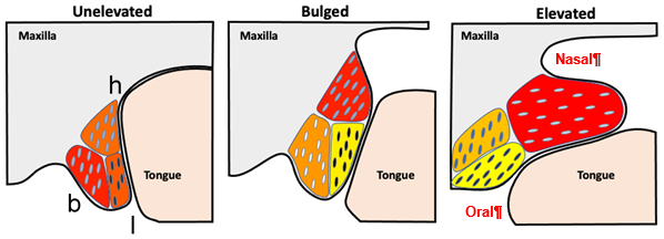
Illustration of palatal shelf elevation process, including cellular changes observed in the buccal (b), lingual (l) and hinge (h) regions of the unelevated, bulged and elevated PS.
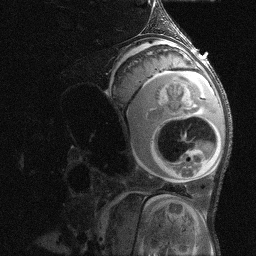
MRI of mouse embryo in utero
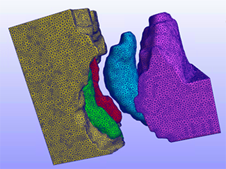
Finite element model from in utero MRI
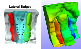
Lateral buldges from MRI and predicted displacements correspond