Distal Radioulnar Contact Mechanics
Student: Mathew Sunil Varre
Investigators: Ken Fischer, PhD; Sang-Pil Lee, PhD; Terence McIff,
PhD; E. Bruce Toby, MD
Distal Radioulnar Joint (DRUJ) is a joint of the wrist which allows forearm rotation and force transmission in the upper limb while preserving stability independent of flexion and extension of the forearm. It is a frequently injured part of the body. Conditions affecting the joint could be positive ulnar variance (Ulnar Impaction Syndrome) or negative ulnar variance (ulnar impingement). It is also adversely affected by nearby injuries such as distal wrist fractures. In fact, significant correlation has been found between negative ulnar variance and scapholunate dissociation [1, 2, 3]. This leads to the question of whether or not scapholunate dissociation causes changes in the radioulnar joint mechanics. Altered joint mechanics are highly associated with onset of secondary osteoarthritis (OA). An understanding of the normal and pathological wrist in vivo radioulnar joint contact mechanics should help physicians make better clinical recommendations and improve treatment for the primary injury and DRUJ pathology. Successful treatment may effectively prevent onset of OA.
The overall goal of our research program is to characterize normal and pathological wrist mechanics during functional loading to evaluate surgical repair and to relate changes in mechanics with pathology to onset of OA. The objective of this study was to evaluate the contact mechanics of DRUJ in wrists with scapholunate dissociation and in contralateral uninjured wrists based on MRI scans during functional loading. We hypothesized that pressures and contact areas would be higher for injured wrists than for controls.
Patients diagnosed with unilateral scapholunate dissociation and no wrist arthritis participated in this study. The MR Imaging was done at Hoglund Brain Imaging Center (HBIC) located at the University of Kansas Medical Center. A 3T Clinical MRI scanner was used in CISS sequence with a custom made coil placed on the wrist joint. Two sets of images were taken one in relaxed state and one with light grasp.
Image registration was used to determine the kinematics from the bone surface models to match the functionally loaded state using ANALYZE 5.0. Models of the bones radius and ulna were constructed using the MR images obtained from the relaxed scans including the cartilage in the models using MPX Image software. Contact analysis was performed using Joint_Model Program [4]. The region of surface penetration yielded the contact area, the local depth of interpenetration was used to estimate local cartilage deformation. Assuming uniform cartilage thickness f 1mm and an effective cartilage modulus of 4MPa, a local contact pressure was calculated from the cartilage strain estimate. Contact pressures were integrated over the contact area to obtain contact force. Contact parameters were compared between normal and injured joints.
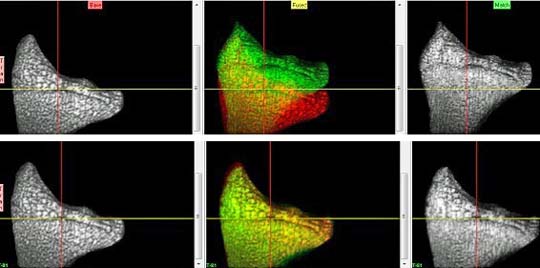
Radius registration as seen in Analyze 5.0 (Mayo Clinic, MN)
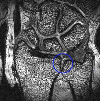
The blue circle encloses part of the Distal Radioulnar Joint
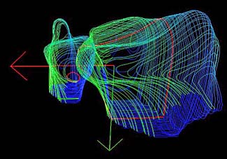
3D Point cloud of Radius and Ulna
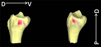
A model showing contact area (in red) on Radius