Mid-Carpal Contact Mechanics
Student: Madhan Sai Kallem
Investigators: Ken Fischer, PhD; Sang-Pil Lee, PhD; Terence McIff,
PhD; E. Bruce Toby, MD
Wrist can be subject to many injuries. Scapholunate (SL) dissociation is a relatively common injury that is particularly difficult to diagnose and treat. Without treatment, SL dissociation is known to progress to scapholunate advance collapse (SLAC wrist) and associated osteoarthritis (OA). Traumatic arthropathy of the wrist due to scapholunate dissociation has a definitive pattern from onset to severe bone and joint degeneration. The altered mechanics and joint wear together with the developed SLAC wrist cause a secondary carpal collapse between the capitate and lunate. The initial SL disruption causes apparent changes in joint kinematics and contact patterns. Thus, understanding normal and abnormal in vivo contact mechanics as a result of SL ligament injury may lead to more effective treatments that may even prevent the onset of OA. In addition, in vivo contact mechanics data after surgical treatment may help determine the effectiveness of various surgical techniques which are used to correct SL injury. The objective of this study was to compare midcarpal joint mechanics of the capitate as it articulates with the scaphoid and lunate in both injured wrists with SL dissociation and in the normal contralateral control wrists before surgical treatment and after the treatment.
The scans for this study were performed using a 3T Siemens Allegra MRI scanner with a custom coil for the wrist. A constructive interface steady state(CISS) sequence was used to perform two sequential scans for each wrist(injured and normal), one with the hand and wrist relaxed and the other during active grasp. High resolution relaxed scans were used to build surface models of capitate, lunate and scaphoid along with their cartilage. The active grasp scans were used to obtain kinematics of the carpal bones from unloaded to the functionally loaded state as determined by the registration (Analyze 5.0). Contact mechanics parameters were calculated using the Joint_Model software. The kinematics from image registration was applied to the surface models to obtain contact areas, contact forces, average contact pressures, and peak contact pressures.
Preliminary results showed adverse changes in contact parameters from uninjured to injured for both the scaphocapitate and lunocapitate articulations. In addition to the contact prameters, the shift in contact location and loading on different areas of cartilage are likely to initiate onset of OA. Figure 3 shows an example of a capitate model showing contact variations between Normal and Injured along different contact interfaces. The study is still going and additional subjects will provide more definitive conclusions
.
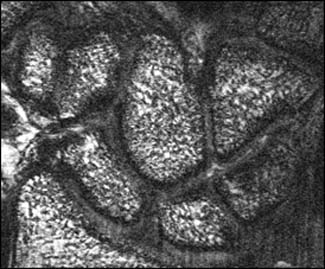
A typical MRI
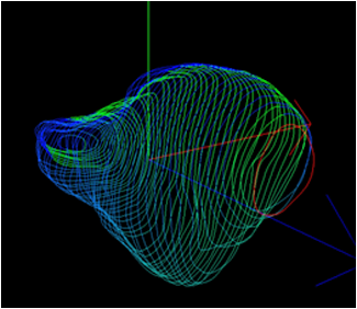
Segmented Capitate
