Development of an Equine Stifle Joint Finite Element Model
Student: Lance Frazer
Investigators: Ken Fischer, Ph.D.; Elizabeth Santschi, DVM
Subchondral bone cysts (SBCs), sometimes referred to as subchondral lesions or subchondral lucencies, continue to plague young horses, especially racehorses. These cystic lesions cause pain (lameness) and severely limit their performance. With 50+ years of research dumped into this problem, there still reamins to be seen both an accepted theory of development (we hypothesize that mechanical trauama and overload are the primary cause), as well as a universally accepted treatment. Current treatments involve SBC debridement and subsequent filling of the void with some combination of biological substances. Unfortauntely, research suggests that full radiographic healing post-surgery is below 20%. Therefore, the primary goal of this study was two-fold. 1) Develop a finite element model of an equine stifle joint to better understand (mechanically) how these cysts initiate and develop, and 2) test and evaluate the efficacy of a novel surgical treatment that shows early promise of a high incidence in radiographic healing.
The first step was to create a model and validate it. This was accomplished through the use of CT images and comparing computational data with experimental data. Once the model was created, we could start to answer an initial question. How does a cyst impact mechanical stresses in the surrounding bone? We ran models of three different cyst sizes (0.03 cm3, 0.5 cm3, and 1 cm3) and found substantial differences in shear and tensile stresses for each cyst size [1].
The next step beyond creating the initial model was to examine how the complexity of different model components, such as meniscal attachments and material properties, affected bone stresses. We found that the complexity of meniscal attachement is critical when assessing tensile stresses and that simplified material properties can be justified if the output variables are well understood [2].
In order to expand our understanding of how a void impacts bone stresses, we applied loads similar to measured ground reaction forces in the forelimb during gallop in models with two different cyst sizes (no cyst and 2 cm3 cyst) and with various degrees of internal femoral rotation. What did we find? Peak tensile, compressive, and shear stresses increased with the presence of a larger void and with internal femoral rotation in both the bone and the medial meniscus. Shear stresses both with and without a void were near an approximated yield strength [3]. This further suggested that mechanical trauma plays a role in the formation and growth of SBCs.
Our focus then shifted towards understanding how the use of lag screws promoted bone growth into the void of the cyst. We found that the presence of a lag screw changed the orientation of stress across a 2 cm3 cyst. This was most noticeable when the screw was placed in a proximodistal oblique orientation [4]. This provided supporting evidence for a successful treatment plan utilized in the veterinary field.
Additionally, we ran simulations to test the effects of lag screw placement on bone stresses and bone formation in various cyst sizes. Our results? We found that larger cysts, which allow lag screw penetration, respond more effectively to this kind of treatment [5].
Next, we asked ourselves another question. Does pressurized fluid within the cyst contribute to its growth? To begin answering this question, we ran two types of models to simulate intra-cyst fluid pressure within three cyst sizes (0.03 cm3, 0.5 cm3, and 1 cm3). The first type was a model that contained a consistent normal pressure of 0 Pa, 1 kPa, 100 kPa, 1 MPa, 3 MPa, 5 MPa, or 10 MPa over the inside of the cyst wall. The second type of model filled the cysts with a solid, assigned properties to represent an incompressible fluid, to represent the internal fluid. Both types of models indicated that internal cyst fluid pressure does not play a substantial role in the growth of cysts (manuscript in process).
Sources
1. Frazer LL, Santschi EM, Fischer KJ: The Impact of Subchondral Bone Cysts on Local Bone Stresses in the Medial Femoral Condyle of the Equine Stifle Joint. Medical Engineering and Physics, 2017;48:158-167 doi: http://dx.doi.org/10.1016/j.medengphy.2017.06.019
2. Frazer LL, Fischer KJ: Evaluating the Effect of Various Meniscal Attachments and Material Properties on Femoral Bone Stress. Journal of Musculoskeletal Research, Vol. 21, No. 1 (2018) 1850003 doi: 10.1142/S0218957718500033
3. Frazer LL, Santschi EM, Fischer KJ. Impact of a Void in the Equine Medial Femoral Condyle on Bone Stresses and Peak Contact Pressures in a Finite Element Model. Vet Surg. 2019;48(2):237-246. doi:10.1111/vsu.13139
4. Frazer LL, Santschi EM, Fischer KJ: Stimulation of Subchondral Bone Cyst Healing by Placement of a Transcondylar Screw in the Equine Medial Femoral Condyle. Veterinary Surgery, 2019, 48(7):1194-1203. doi:10.1111/vsu.13247
5. Frazer L, Santschi EM, Ring SJ, Hewitt RE, Fischer K. Impact of Size and Shape of Equine Femoral Subchondral Bone Cysts with a Transcondylar Screw On Predicted Bone Formation Area in a Finite Element Model [published online ahead of print, 2020 Jan 1]. J Biomech Eng. 2020;10.1115/1.4045892. doi:10.1115/1.4045892
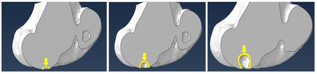
Fig 4: From left to right: 0.03 cm3, 0.5 cm3, and 2 cm cysts3
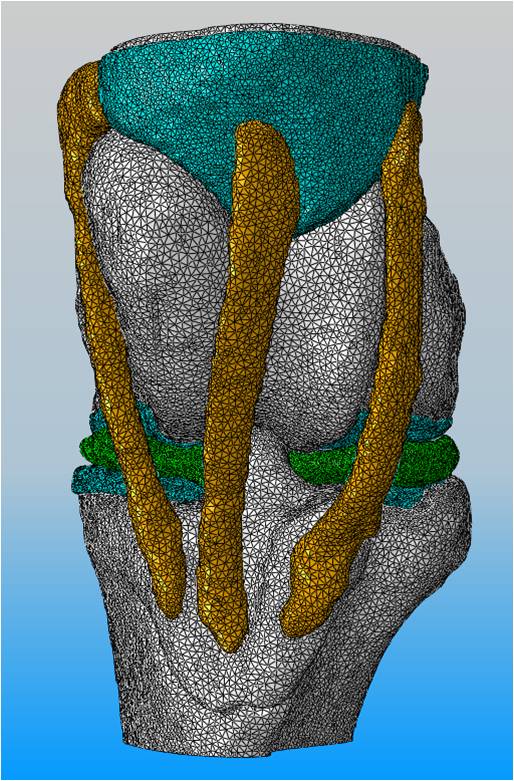
Fig 1: Finite Element Mesh (Anterior View) of an Equine Stifle Joint
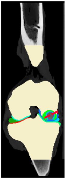
Fig 2: Segmented CT Image with Lag Screw (in red)
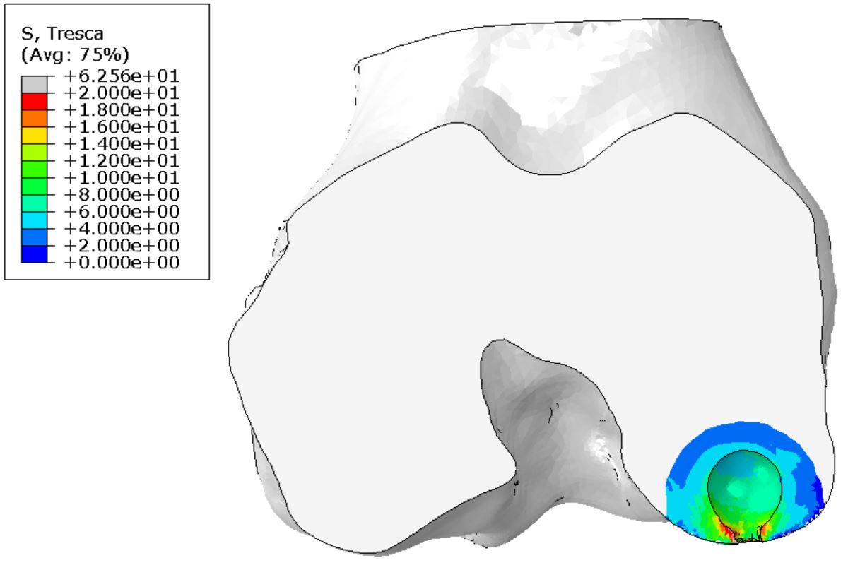
Fig 3: Shear stresses (in MPa) in a region of interest surrounding a 1 cm3 cyst