Radiocarpal Contact Mechanics
Student: Joshua Johnson
Investigators: Ken Fischer, PhD; Sang-Pil Lee, PhD; Terence McIff,
PhD; E. Bruce Toby, MD
Scapholunate dissociation (SL ligament disruption) due to trauma can cause changes in joint kinematics and contact patterns, which can lead to secondary radiocarpal osteoarthritis (OA) with advanced collapse (SLAC wrist). The relationship between consequent abnormal mechanics and the onset of OA is not clearly understood, however elevated joint contact pressure is believed to be an associated factor. Knowing how injuries affect joint physiology and mechanics and how well surgical repairs restore the mechanics may improve surgical efficacy and help predict OA risk. The objective of this research is to compare radiocarpal joint mechanics from injured wrists to contralateral controls and also injured versus surgically repaired wrists using MRI-based contact modeling primarily. Finite element (FE) modeling is also used to investigate 3D stresses and strains from cartilage surface to subchondral bone.
Patients diagnosed with unilateral scapholunate dissociation at the University of Kansas Medical Center are enrolled for participation in the study. MR images are acquired at the Hoglund Brain Imaging Center using a 3T clinical scanner with a custom coil. Two sets of images are obtained for the uninjured and injured wrists; one high resolution set with the hand relaxed to obtain model geometry, and another during active grasp at half the resolution to determine kinematics during functional loading.
To construct models of the radiocarpal joint, the radius, lunate and scaphoid bones (cartilage included) are segmented from the high resolution images to obtain 3D point clouds. To construct surface models, triangular facets are wrapped onto the point clouds to create surfaces for the bones using Geomagic. To construct FE models, cartilage and bone geometry are individually meshed in HyperMesh. The radius, scaphoid, and lunate bones (without cartilage) from the relaxed and active grasp image sets, are isolated on a black background for image registration. Image registration is performed using Analyze 5.0 to determine carpal bone kinematics during functional loading.
Kinematics are implemented with the bone surface contact models in the Joint_Model software and with the FE models in Abaqus to obtain contact forces, contact areas, peak and average contact pressures. Model accuracy is verified by direct contact area measurements acquired from the active grasp scans. Contact parameters together with functional data (pain levels and grip strength) can help us better understand the pathomechanics of injury and the efficacy of surgical reconstruction. Preliminary results show a lateral shift in scaphoid contact which is usually observed with SL dissociation. Also, peak and average pressure appear to be higher in the injured wrist compared to normal indicating detrimental alterations to radiocarpal mechanics as a result of SL injury.

Scaphoid (left) and Lunate (right) contact intensities on the Radius fossa.

Radiograph of SL Dissociation (left); High Resolution MR Image of the Radiocarpal Joint (right)
Segmented 3D Point Cloud (left); FE Mesh of the Radius and Articular Surface (right)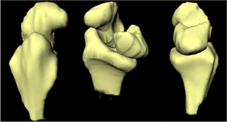
Lateral, Oblique and Medial Views of Radiocarpal Surface Models