Biochemical Assessment of Cartilage
Student: Dannica Sturgeon
Investigators: Ken Fischer, PhD; Sang-Pil Lee, PhD; Terence McIff,
PhD; E. Bruce Toby, MD
The objective of this study is to evaluate changes in the status of the cartilage in the wrist following scapholunate ligament injury by analyzing the T2 relaxation time in the cartilage of human subjects. The T2 relaxation time increases with tissue hydration and is sensitive to biochemical composition, changes in which are markers for cartilage degeneration1. Thus, our hypothesis is that the calculated T2 relaxation time will be higher in the cartilage of injured wrists than in that of contralateral (uninjured) controls. Special images for calculating the T2 time are taken during the scanning of each subject for both their control and injured wrist.
There are four image sets obtained from the spin echo scans, which are first registered to minimize the effects of any motion occurring over the set of 4 scans. The regions of radiocarpal cartilage on the radius and lunate bones in the wrist are then defined manually on the third image set. After this, the data is analyzed by a custom Matlab code to calculate the T2 time for each pixel of cartilage via regression to an exponential decay curve. Descriptive statistics are calculated for the image set, and the mean T2 times for each bone are compared between the control and injured wrists.
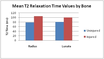
Summary of first 7 subjects analyzed
1. Blumenkrantz, G. et. al., 2007. European Cells & Materials 13, 75-86.
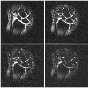
Example set of four images. Notice the decay in intensity throughout the set of four images.
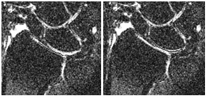
Example of radius (left) and lunate (right) template for this set of images (zoomed in)